Blog
Histoplasmosis
What is histoplasmosis?
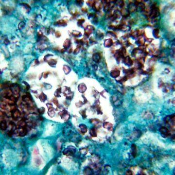
Histoplasmosis is a fungal infection caused by Histoplasma capsulatum (histo) spores, which are inhaled into the lungs. The fungus is found in the soil and may be released into the air when plowing fields, digging holes, or sweeping chicken coops. Histoplasmosis may later cause a serious eye disease called “ocular histoplasmosis syndrome” (OHS), a leading cause of vision loss in Americans between the ages of 20 and 40—primarily in a region of central and southern states known as the “Histo belt.” In those areas, up to 90 percent of adults have been infected with histoplasmosis.
How does ocular histoplasmosis syndrome develop?
While no traces of the histo fungus have been found in the eyes of people with OHS, it is believed that histo spores from the lungs make their way to the eyes and lodge in the choroid, a layer of blood vessels that supply the retina. Fragile, abnormal blood vessels begin to grow under the retina, forming a lesion called choroidal neovascularization (CNV). If left untreated, CNV can become scar tissue, replacing normal tissue in the central part of the retina (the macula). If the abnormal blood vessels grow into a tiny depression in the macular called the fovea, they can cause damage that can severely harm and even destroy central vision.
What are the symptoms of ocular histoplasmosis?
In its early stages, OHS may cause no symptoms, and it usually goes away on its own. So you may never know you had it. However, tiny scars on the retina called “histo spots” may remain and may cause complications months or years later. If this happens:
- Straight lines may appear crooked or wavy
- A blind spot may appear in your central vision
How is ocular histoplasmosis detected?
If you have ever had histoplasmosis, it’s important to see an eye care professional to determine if it has affected your eyes. The best way to learn if you have ocular histoplasmosis is through a comprehensive dilated eye exam. The eye doctor will look for evidence of histo spots and swelling of the retina and examine the blood vessels of your retinas.
Can ocular histoplasmosis be treated?
Treatments for ocular histoplasmosis focus on preventing future vision loss.
- Photocoagulation. This type of laser surgery can reduce the chance of further CNV growth or vision loss. The doctor uses a small, powerful light beam to destroy abnormal blood vessels as well as some overlying retinal tissue. Photocoagulation has been shown to reduce future vision loss from OHS by more than half. Sometimes the abnormal blood vessels grow back, and when this happens, additional laser surgery may be necessary.
- Injections. Therapies that target a protein called “vascular endothelial growth factor” (VEGF)—such as aflibercept or ranibizumab—inhibit abnormal blood vessel growth. These treatments are injected into the eye. Anti-VEGF therapies can slow vision loss and even improve vision in some people. Repeated treatments are often needed to achieve the most benefit.
If you have vision loss from OHS, there are low vision aids you can use to maximize the use of your remaining vision. Examples include handheld lenses, electronic reading machines, and closed circuit video magnification systems.
Source: The National Eye Institute (NEI)